Animal Cell Diagram: A Simple, Clear Guide to the Building Blocks of Life

Have you ever wondered what’s happening inside your body right now—inside your skin, your muscles, even your thoughts? All of it traces back to tiny living units called cells. And when we talk about animals (including humans), it all begins with understanding the structure of an animal cell. This guide walks you through the animal cell diagram in a way that feels natural, easy, and genuinely interesting—no lab coat required.
Understanding Cells: The Basics of Life
Before jumping into diagrams, let’s take a step back. A cell is the smallest unit of life. Think of it as a miniature city where thousands of tasks happen every second. Some cells are simple, others more complex. Animal cells fall into the complex category because they contain many specialized parts working together.
Why does this matter? Because everything your body does—breathing, healing, moving, thinking—depends on how these tiny units function. Learning about their structure helps us understand life itself.
What Is an Animal Cell?
An animal cell is a type of cell found in animals, including humans. Unlike plant cells, animal cells don’t have a rigid outer wall or structures for photosynthesis. Instead, they’re flexible, adaptable, and designed to support movement, communication, and growth.
When you look at an animal cell diagram, you’re essentially seeing a map of all the internal parts that keep the cell alive and active.
Why Diagrams Matter in Learning Biology
Ever tried assembling furniture without instructions? A diagram works the same way—it shows how everything fits together. An animal cell diagram visually explains where each component sits and how it relates to the others.
For students, diagrams make learning easier. For general readers, they turn abstract ideas into something concrete and memorable. Seeing is understanding.
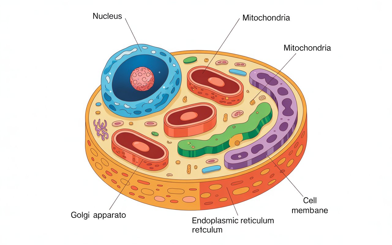
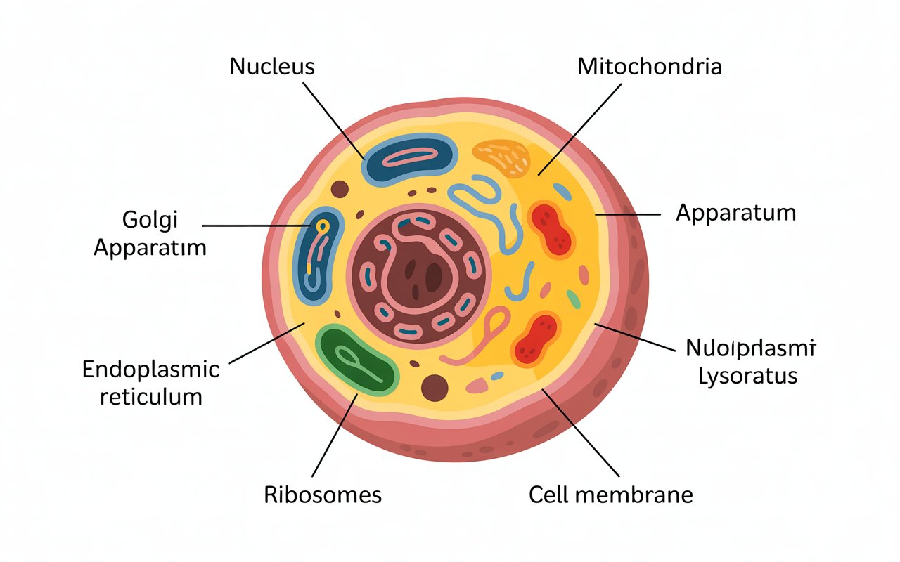
The Cell Membrane: The Protective Gatekeeper
What does it do?
The cell membrane is the thin outer layer that surrounds the cell. It controls what enters and exits, much like a security gate at the entrance of a building.
This structure is flexible, not rigid. That flexibility allows animal cells to change shape when needed—essential for processes like movement and division. It also helps maintain balance by allowing nutrients in and waste out.
Cytoplasm: The Cell’s Living Space
Inside the membrane lies the cytoplasm, a jelly-like substance that fills the cell. This is where most cellular activities occur.
Imagine the cytoplasm as the floor space of a factory. All the machines (cell parts) are placed here, and materials move around freely. Without this medium, nothing inside the cell could interact effectively.
The Nucleus: The Control Center
Why is it important?
The nucleus acts like the brain of the cell. It stores genetic information and directs all activities, from growth to repair.
Inside the nucleus, you’ll find DNA, which carries instructions that make every organism unique. If the cell were a company, the nucleus would be the CEO’s office—making decisions and issuing commands.
Mitochondria: The Power Producers
Mitochondria are often called the powerhouse of the cell, and for good reason. They generate energy that the cell needs to function.
Think of them as tiny power plants. The more active a cell is, the more mitochondria it contains. Muscle cells, for example, have a lot because they require constant energy.
Ribosomes: The Protein Builders
Ribosomes are small but mighty. Their job is to assemble proteins, which are essential for structure, repair, and communication within the body.
These structures can float freely in the cytoplasm or attach to other components. Either way, they’re like skilled workers assembling products on a production line.
Endoplasmic Reticulum: The Transport Network
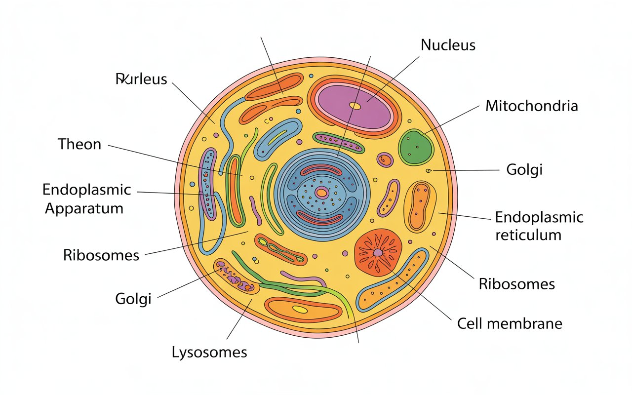
The endoplasmic reticulum (ER) is a network of folded membranes that helps move materials around the cell.
- Rough ER has ribosomes attached and helps process proteins.
- Smooth ER lacks ribosomes and plays a role in making lipids and detoxifying substances.
Picture it as the cell’s highway system, ensuring materials reach the right place efficiently.
Golgi Apparatus: The Packaging Center
Once proteins and other materials are made, they need sorting and shipping. That’s where the Golgi apparatus comes in.
It modifies, packages, and sends products to their destination. If the cell were a post office, this would be the sorting and delivery department.
Lysosomes: The Cleanup Crew
Cells generate waste, just like we do. Lysosomes handle that problem by breaking down unwanted materials and recycling useful parts.
They’re the janitors of the cell—cleaning up debris and keeping everything running smoothly. Without them, waste would pile up and disrupt cell function.
Centrosome and Cytoskeleton: Shape and Support
The centrosome helps organize cell division, while the cytoskeleton provides structure and shape.
Together, they act like scaffolding and planning tools, ensuring the cell maintains its form and divides properly when needed.
Animal Cell vs. Plant Cell: Key Differences
Animal cells and plant cells share many features, but there are important differences. Plant cells have a rigid wall, chloroplasts, and large storage spaces. Animal cells lack these but make up for it with flexibility and specialized functions.
Understanding these differences helps explain why plants and animals live and behave so differently.
How to Read and Label an Animal Cell Diagram
Looking at a diagram can feel overwhelming at first. Start by identifying the outer boundary, then move inward. Focus on one part at a time and think about its role.
Ask yourself: What would happen if this part didn’t work? That question alone can deepen your understanding dramatically.
Common Misconceptions About Animal Cells
Many people think cells are static or simple. In reality, they’re dynamic and incredibly complex. Another common misunderstanding is that all cells look the same. In fact, shape and structure vary depending on function.
Clearing up these myths helps us appreciate just how remarkable life really is.
Conclusion: Why Animal Cell Diagrams Matter
Understanding an animal cell diagram is like learning the blueprint of life. Each part has a purpose, and together they create a system capable of astonishing complexity. Whether you’re a student, a curious reader, or someone brushing up on basics, this knowledge builds a deeper appreciation for how living beings function at the smallest level.
Cells may be tiny, but their impact is enormous.
Frequently Asked Questions
What is the main purpose of an animal cell diagram?
It helps visualize and understand the structure and function of different cell components clearly.
How is an animal cell different from a plant cell?
Animal cells lack a rigid wall and photosynthetic structures, making them more flexible in shape.
Why is the nucleus important in a cell?
It stores genetic information and controls all major cellular activities.
What happens if mitochondria stop working?
The cell would lose its energy supply and eventually fail to function.
Are animal cells visible to the naked eye?
No, they are microscopic and require a microscope to be seen clearly.


Post Comment